This paired triangle contains some very important structures such as the common carotid artery internal carotid.
Floor muscles of digastric triangle.
What are the contents of the submandibular triangle.
The digastric muscle is composed of two bellies anterior and posterior connected by an intermediate round tendon.
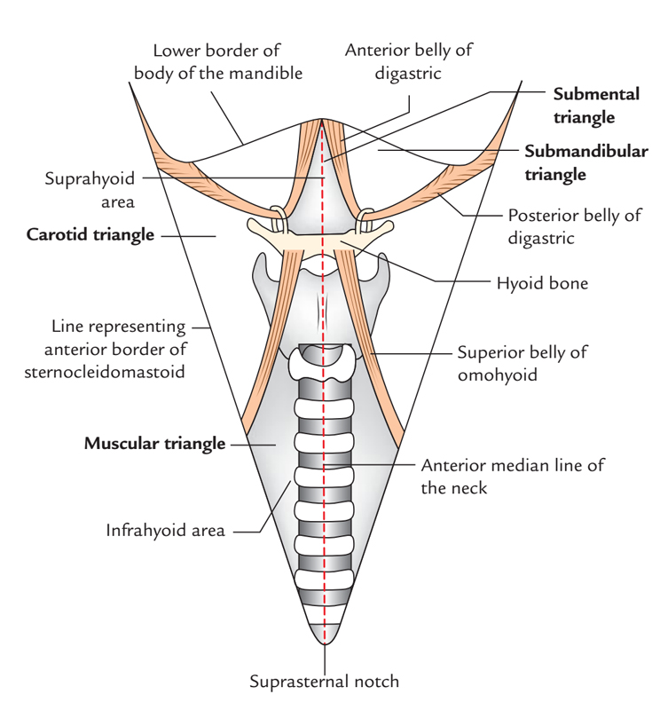
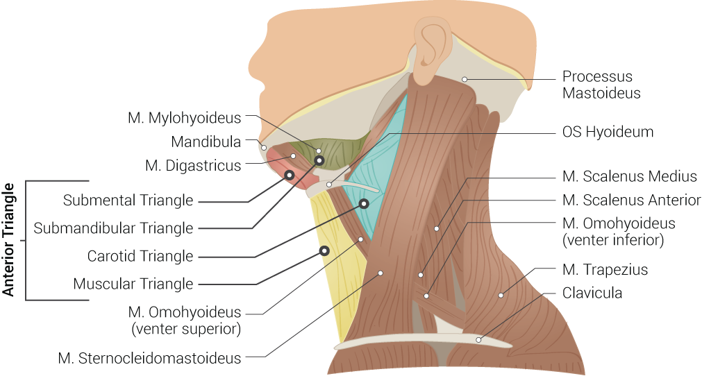
The anterior triangle is subdivided by the hyoid bone suprahyoid and infrahyoid muscles into four triangles.
The carotid triangle of the neck has the following boundaries.
The digastric muscle also digastricus named digastric as it has two bellies is a small muscle located under the jaw the term digastric muscle refers to this specific muscle.
Below by the posterior belly of the digastricus.
Suprahyoid muscles digastric ant and post belly mylohyoid geniohyoid and stylohyoid.
Superior posterior belly of the digastric muscle.
A major landmark of the submandibular triangle is the submandibular gland innervated by the facial nerve.
However other muscles that have two separate muscle bellies include the ligament of treitz omohyoid occipitofrontalis.
The two bellies of the muscle have different embryonic origins and hence are supplied by different cranial nerves.
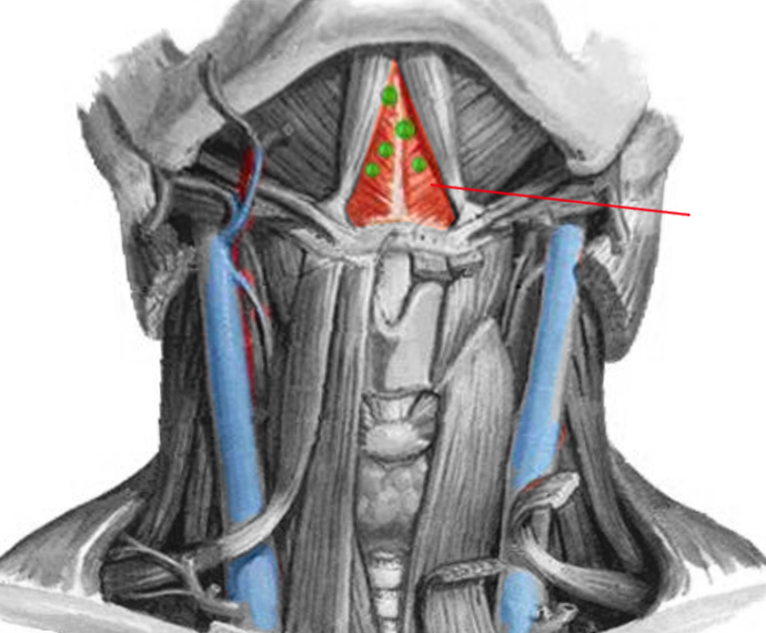
On floor of mouth between mandible and genioglossus.
Muscles nerves blood vessels glands.
The carotid triangle the submental triangle and the submandibular triangle.
Investing fascia covers the roof of the triangle while visceral fascia covers the floor.
Digastric or submandibular triangle.
Submandibular triangle is bordered by the mandible and bellies of the digastric muscle.
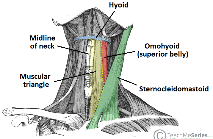
Infrahyoid muscles omohyoid sternohyoid sternothyroid and thyrohyoid.
It lies below the body of the mandible and extends in a curved form from the mastoid.
2 bellies of digastric anterior.
Mastoid process of temporal bone.
Muscles digastric muscle stylohyoid geniohyoid mylohyoid hyoglossus middle pharyngeal constrictor nerves mylohyoid nerve cn v.
The posterior belly of digastric muscle forms the superior border of the carotid triangle.
It is covered by the integument superficial fascia platysma and deep fascia ramifying in which are branches of the facial nerve and.
In front by the anterior belly of the digastricus.
Digastric triangle also known as submandibular triangle is named due to it is position in the middle of the two bellies of the digastric muscle and inferior towards the base of the mandible.
Lateral medial border of the sternocleidomastoid muscle.
This salivary gland can be described as having two lobes which are divided by the posterior border of the mylohyoid muscle.
Digastric fossa on the deep surface of symphysis menti of the mandible.
The digastric muscle divides the anterior triangle of the neck into three smaller divisions.