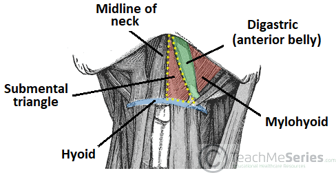
Chin is the apex h.
Floor of carotid triangle diagram.
Alexandra sieroslawska md reviewer.
Medially sagittal line down the midline of the neck.
This triangle is situated between the ophthalmic and maxillary divisions of the trigeminal nerve and the bone of the middle fossa between the foramen rotundum and superior orbital fissure figs.
Francesca salvador msc last reviewed.
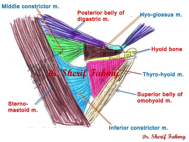
Investing fascia covers the roof of the triangle while visceral fascia covers the floor.
Quizzes labeled diagrams and articles.
Superior belly of omohyoid m.
Hyoid bone is the base in.
Anterior triangle submental triangle mnemonic.
Posterior belly of digastric m.
In between digastrics sides digastric triangle mnemonic.
Hyoid bone thyro hyoid m.
The carotid triangle or superior carotid triangle is a portion of the anterior triangle of the neck coverings and boundaries.
Thus it is an area of both anatomical and clinical importance.
Floor formed by prevertebral fascia covering underlying muscles splenius capitis levator scapulae posterior middle and anterior scalene further divided by inferior belly of the omohyoid anterior cervical triangle boundaries.
A gland located beneath the floor of the mouth.
The anterior triangle is situated at the front of the neck.
Constrictores pharyngis medius and inferior.
Posteriorly by the.
Laterally anterior border of the sternocleidomastoid.
Triangles of the neck diagram.
Its floor is formed by parts of the thyrohyoid membrane hyoglossus and the.
This space is used to expose the superior orbital vein and the sixth cranial nerve and to access carotid cavernous fistulae.
Contents of carotid triangle dr.
Triangles of the neck.
In this article we shall look at the borders contents and clinical correlations of the femoral triangle.
Carotid triangle submandibular triangle submental triangle.
It is comprised of various anatomical structures that are noted as theoretical borders.
The roof is formed from the skin fascia and platysma.
Borders and contents of the supraclavicular.
Floor of carotid triangle dr.
Many large neurovascular structures pass through this area and can be accessed relatively easily.
Mastoid and mandible is base.
The first digitation of serratus anterior and the first rib are in the floor of this triangle.
Superiorly inferior border of the mandible jawbone.
The facial artery arises from the external carotid artery s carotid triangle and it travels a course passing the lingual artery.