Floor Of Left Maxillary Sinus
Panoramic Radiograph Of The Case Opacification Of Left Maxillary Download Scientific Diagram
Benign Maxillary Sinus Masses Ento Key

Cbct Images Of Left Maxillary Sinus In Sagittal View Depicting Relation Download Scientific Diagram

Thick Inspissated Mucous Covering The Left Maxillary Sinus Floor Download Scientific Diagram
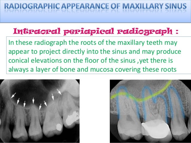
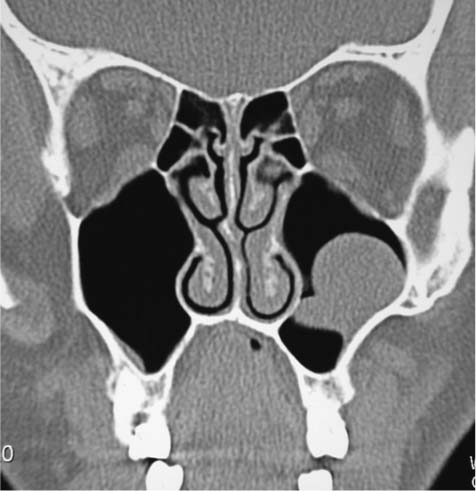
Radiology Of Maxillary Sinus

Isolated Unilateral Upper Alveolar Numbness In Silent Sinus Syndrome Bmj Case Reports

These cavities are called sinuses and they are located in the maxilla or upper jaw cysts are closed pocket like formations of tissue and are filled with liquid air or semi solid material.
Floor of left maxillary sinus.

Postoperative Stage 2 Panoramic Radiograph Of Left Maxillary First Download Scientific Diagram

Cranio Facial Ct Scan Coronal Sections Left Maxillary Sinus With Download Scientific Diagram

Pns View Showing Destruction Of Floor Of Maxillary Antrum On Left Side Download Scientific Diagram

Intraoperative Endoscopic Views Of The Left Maxillary Sinus Of An Orbital Floor Fracture Through The Antral Window A The Orbital Floor Was Fractured And Periorbital Soft Tissue Was Herniated Into The Maxillary
Source : pinterest.com
