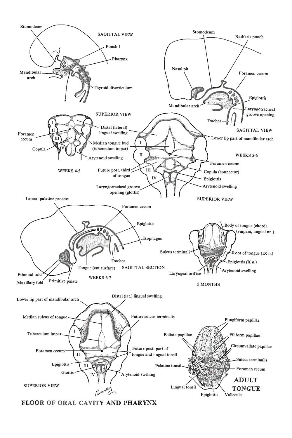
Floor of the pharynx development of the tongue.
Floor of the pharynx.
Size of opening is controlled circumoral muscles such as orbicularis oris buccinator depressors and.
The upper floor the nasopharynx is primarily a passageway for air and secretions from the nose to the oral pharynx.
The nasopharynx oropharynx and laryngopharynx from superior to inferior.
Thick fibres of muscle and connective tissue attach the pharynx to the base of the skull and surrounding structures.
The bottom of your mouth located under your tongue is called the floor.
1 oral vestibule 2 oral cavity proper oral vestibule slit space between teeth buccal gingiva and lips cheek.
Glossopharyngeal vagus and sympathetic nerves.
Floor of the pharynx 1.
Oral cavity 2 parts.
The pharynx is a wide short tube with muscular dorsal and lateral walls.
But there are secondary functions.
Floor of oral pharynx under the mucus membrane covering the root of the tongue.
The muscles of these walls are inserted latero ventrally into the hyoid bone and the cartilages of the larynx.
For the anatomical description the pharynx can be divided into three floors.
How is the pharyngeal plexuses formed.
The middle pharyngeal constrictor is located on the lateral and posterior sides of the neck it is found anterior to the prevertebral muscles such as longus coli and longus capitis and posterior to the muscles of the mouth floor most notably the hyoglossus muscle.
Development of thyroid gland.
Floor of the pharynx 2.
It is also connected to the tympanic cavity of the middle ear through the auditory tubes that open on both lateral walls.
The act of swallowing opens briefly the normally collapsed.
It acts as the first line of defense.
Anatomy of oral cavity and pharynx dr mohit goel jr1 22 aug.
Both circular and longitudinal muscles occur in the walls of the pharynx.
Throat cone shaped passageway leading from the oral and nasal cavities in the head to the esophagus and larynx the pharynx chamber serves both respiratory and digestive functions.
Why are the tonsil arranged in a ring form.
It is comprised of three parts.
The pharynx is a muscular tube that connects the nasal cavities to the larynx and oesophagus.
The primary function of the oral cavity is firstly the selection of food via taste yes but also of course via smell and sight.
The functions of the oral cavity.
Vestibule communicates with exterior through mouth.
It is common to both the alimentary and the respiratory tract.
Tongue largest single muscular organ in oral cavity.
The nasopharynx the oropharynx and the laryngopharynx.
The oral mucosa are the tissues that line the interior of your mouth while the salivary glands produce saliva.
Having selected the food then the mouth ingests the food takes the food in masticates the food chews it and swallows the food so that it goes down to the rest of the digestive tract.
Within the pharynx the middle pharyngeal constrictor sits between the superior and inferior pharyngeal constrictor muscles.
The pharynx or throat is a tube about five inches long composed of three parts.
The tube begins at the base of the skull and ends inferior to the cricoid cartilage c6.
7th 9th 10th cranial nerves.
The floor is provided by the soft palate.
Oral cavity pharynx radio anatomy 1.