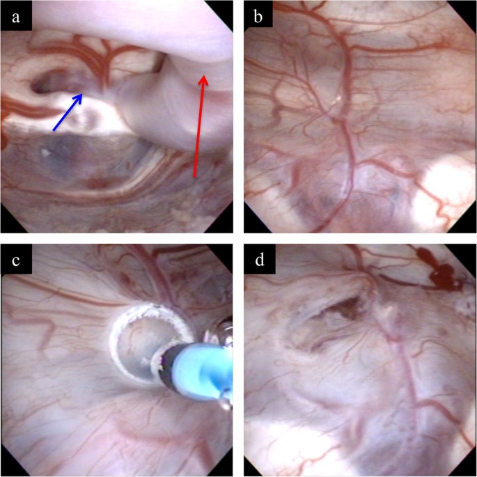
The floor of third ventricle is perforated anterior to the halfway point between infundibular recess and mamillary bodies and a balloon dilatation technique is the most used during the ventriculostomy.
Floor of third ventricle perferated.
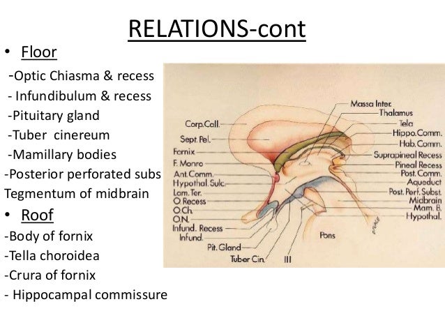
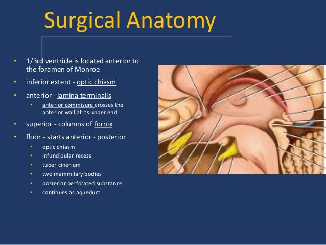
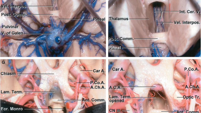
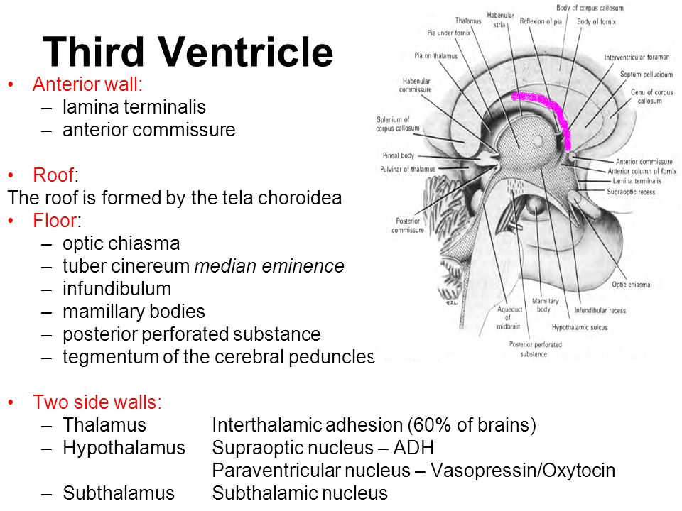
Floor and roof the floor is formed by the optic chiasma the tuber cinereum and the infundibulum the mamillary bodies the posterior perforated substance and the tegmentum of the midbrain.
The scope was then removed the craniostomy plugged with gel foam and a layered closure was subsequently performed.
39 40 the etv procedure consists of advancing a fiberoptic endoscope into the lateral ventricle through the foramen of monro into the third ventricle so that under direct visualization the membranous floor of the third ventricle can be perforated bluntly followed by enlargement of the fenestration using a balloon.
An endoscopic third ventriculostomy can be performed in order to release extra fluid caused by hydrocephalus.
Floor it is actually the floor of third ventricle it extends across optic chiasma tuber cinerium infundibulum and mamillary bodies to posterior perforated substance 6.
Its upper part assists in forming the floor of the third ventricle.
Like other ventricles the third ventricle has a cavity an anterior wall a posterior wall a floor a roof and two lateral walls.
The floor of the third ventricle is formed by a number of structures including the hypothalamus subthalamus mammilary bodies infundibulum pituitary stalk and the tectum of the midbrain.
These cells produce cerebrospinal fluid.
The posterior perforated substance is the depressed area between the crura is termed the interpeduncular fossa and consists of a layer of gray matter which is pierced by small apertures for the transmission of blood vessels.
Patients were stratified into three groups.
As a general rule etv is used for obstructive rather than communicating hydrocephalus 37 38 although its use has been reported in ah.
The floor of the third ventricle is formed by hypothalamic structures and this can be opened surgically between the mamillary bodies and the pituitary gland in a procedure called an endoscopic third ventriculostomy.
This procedure can cause a variety of complications reported in the literature.
Its lower part lies on the ventral aspect of the medial portions of the tegmenta and contains a nucleus named the interpeduncular ganglion.
37 while in theory this is a simple.
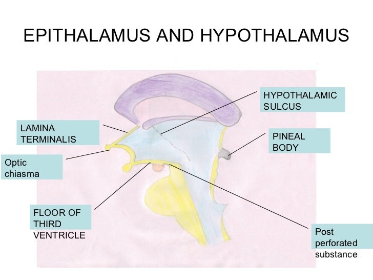
It is a cavity within diencephalon it is a midline slit like cavity situated between the two thalami and the part of hypothalamus.
Side walls these are the lateral walls of third ventricle formed by thalamus hypothalamic groove or sulcus and hypothalamus itself.
A post operative mri shows very small residual under the corpus callosum adherent to the roof of the left lateral ventricle.
The floor of the third ventricle was then perforated and dilated with a four french fogarty catheter bipolar cautery and irrigation were used as necessary for hemostasis.
The lateral walls of the third ventricle are formed by the walls of the left and right thalamus.
Describe the location and boundaries of third ventricle.