This procedure can cause a variety of complications reported in the literature.
Floor of third ventricle perforation.
The floor of third ventricle is perforated anterior to the halfway point between infundibular recess and mamillary bodies and a balloon dilatation technique is the most used during the ventriculostomy.
Short term memory loss is another potential complication of endoscopic third ventriculostomy.
The ventricular floor can lead to bleeding as can damage to ventricular walls or perforation of the basilar artery.
The third ventricle is a narrow cavity located between the two hemispheres of the diencephalon of the forebrain the third ventricle is part of a network of linked cavities cerebral ventricles in the brain that extend to form the central canal of the spinal cord the cerebral ventricles consist of the lateral ventricles third ventricle and fourth ventricle.
The floor of the third ventricle was subsequently fenestrated in a standard fashion often with balloon dilation through the endoscope.
Transient diabetes insipidus one of its rarest complications.
The basilar artery can also be seen between the mammillary arteries and must be avoided upon perforation of the third ventricular floor.
Running through the third ventricle is the interthalamic adhesion which contains thalamic.
The floor of the third ventricle was then perforated and dilated with a four french fogarty catheter bipolar cautery and irrigation were used as necessary for hemostasis.
The scope was then removed the craniostomy plugged with gel foam and a layered closure was subsequently performed.
The third ventricle is one of the four connected ventricles of the ventricular system within the mammalian brain it is a slit like cavity formed in the diencephalon between the two thalami in the midline between the right and left lateral ventricles and is filled with cerebrospinal fluid csf.
B view of the floor of the third ventricle.
Large bleeds due to vessel injury under the third ventricle can be catastrophic but they are rare.
Etv is technically difficult in post infective hydrocephalus especially in acute phase of disease due to presence of inflammation thick and opaque floor of third ventricle 7 10 17 it is comparatively simple in chronic phase of disease there is an increased risk of hemorrhage and neurovascular injury especially in acute phase.
The endoscope is then advanced into the third ventricle and the floor of the third ventricle is confirmed in the midline anterior to the mammillary bodies and posterior to the infundibular recess fig.
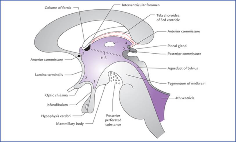
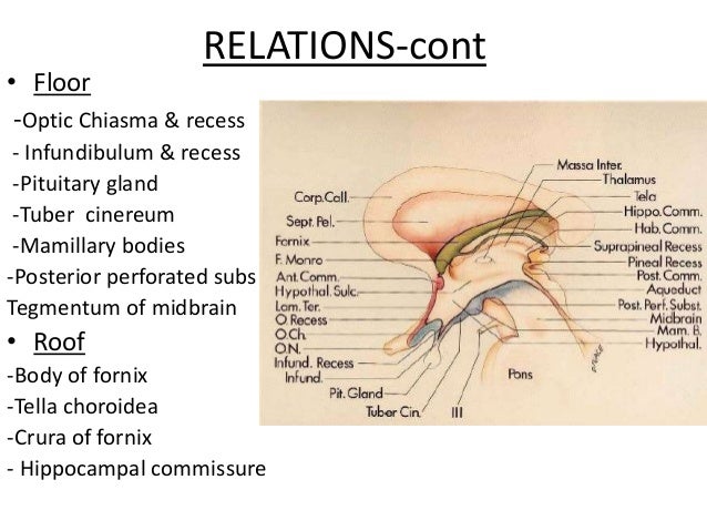
Floor and roof the floor is formed by the optic chiasma the tuber cinereum and the infundibulum the mamillary bodies the posterior perforated substance and the tegmentum of the midbrain.
Like other ventricles the third ventricle has a cavity an anterior wall a posterior wall a floor a roof and two lateral walls.